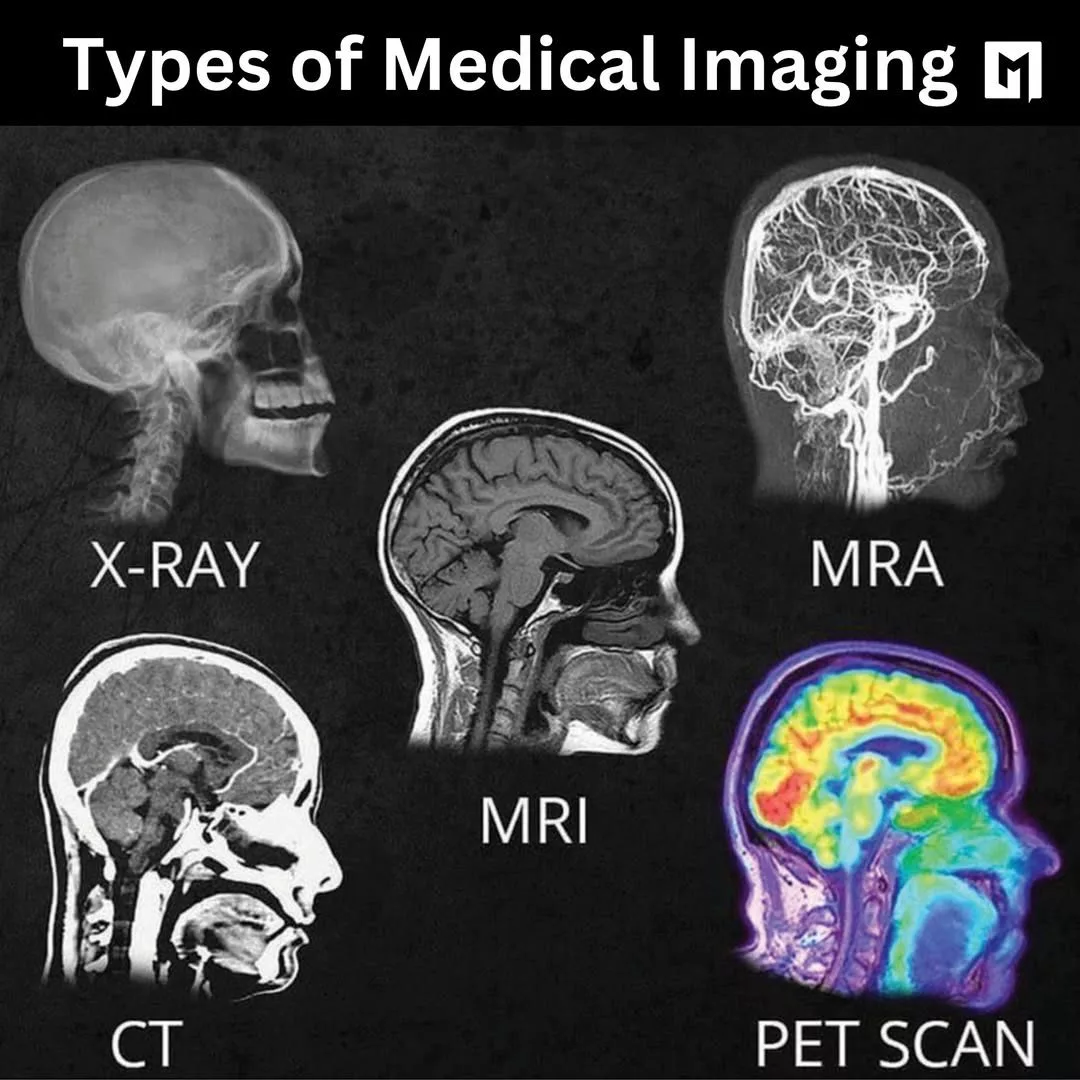
What’s the difference between X-Ray, CT Scan, MRI, and more?
X-Ray – Uses electromagnetic radiation to capture images of bones and dense structures within the body. It is commonly used to detect fractures, infections, tumors, and other abnormalities. X-rays are fast, relatively affordable, and widely available.
CT Scan (Computed Tomography) – Combines X-rays with advanced computer processing to generate detailed cross-sectional images of the body. Unlike traditional X-rays, CT scans visualize bones, organs, blood vessels, and soft tissues, making them effective for diagnosing internal injuries, tumors, infections, and blood clots.
MRI (Magnetic Resonance Imaging) – Utilizes a strong magnetic field and radio waves to produce highly detailed images of soft tissues, organs, and internal structures. It provides excellent contrast between tissue types and is particularly valuable for imaging the brain, spinal cord, joints, and muscles. Since MRI does not use ionizing radiation, it is a safer option, especially for pregnant women and children. However, MRI scans take longer, are more expensive, and may not be suitable for individuals with certain metal implants or medical devices.
PET Scan (Positron Emission Tomography) – Measures metabolic activity in different areas of the body, particularly by detecting glucose (sugar) consumption. Since cancer cells consume large amounts of sugar to fuel rapid growth, they appear as the brightest areas on a PET scan. This imaging technique is commonly used to detect cancerous cells and evaluate their spread.
MRA (Magnetic Resonance Angiography) – A specialized MRI technique that visualizes blood flow within the brain’s vascular system. It is used to detect narrowing, blockages, or abnormalities in blood circulation.